
Arvind Saini, MD, Builds His Practice and Relationship with Topcon
For more than 90 years, Topcon has mastered the art of outstanding customer service. Watch as Ophthalmologist Arvind Saini shares his experience purchasing directly through Topcon and experiencing that service firsthand.
Topcon Healthcare Exceeds 20,000 Maestro OCTs Sold Worldwide
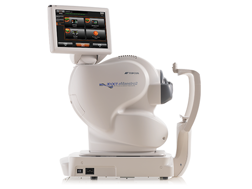
Topcon Healthcare announced today that it has surpassed 20,000 units of sale globally of its Maestro Robotic OCT.
Kerry Salsberg, OD, Shares How Topcon Technology Elevates Patient Care
Watch as Kerry Salsberg, OD, from Eyes On Sheppard, a leading eye care practice in Toronto, shares his experience with Topcon technology and how it has transformed their approach to patient care
IOTM: Choroidal Nevus with Drusen
This 52 year-old patient presented with a sudden central visual field defect in the right eye for two days. Visual acuity, intraocular pressure and anterior segment were unremarkable. Visual field testing revealed a horizontal circumscribed defect from the optic nerve, beyond the macula, inferior to the fovea. (Image A) Fundus photography demonstrated an occlusion of a cilioretinal artery (branch retinal artery occlusion). (Image B) The horizontal SS-OCT (Image C) and vertical SS-OCT (Image D) through the branch retinal artery occlusion clearly demonstrate retinal ischemia.
IOTM: Branch Retinal Artery Occlusion
This 52 year-old patient presented with a sudden central visual field defect in the right eye for two days. Visual acuity, intraocular pressure and anterior segment were unremarkable. Visual field testing revealed a horizontal circumscribed defect from the optic nerve, beyond the macula, inferior to the fovea. (Image A) Fundus photography demonstrated an occlusion of a cilioretinal artery (branch retinal artery occlusion). (Image B) The horizontal SS-OCT (Image C) and vertical SS-OCT (Image D) through the branch retinal artery occlusion clearly demonstrate retinal ischemia.
IOTM: Toxoplasmosis Retinochoroiditis
A 57-year-old Indian female with a prior history of posterior uveitis was referred for scarring OD. BCVA 20/20 OU, diagnostic testing demonstrated +IgG antibodies to toxoplasmosis, IgM was negative.
From Cold Start to Cashflow: Tips for Growing a New Practice
It took Kevin Semma, OD, about a year to move from the idea of opening a new practice to actually opening the doors at Maple Eye Care and Eyewear in Bloomfield Hills, Michigan.
IOTM: Optic Nerve Head Drusen (ONHD)
This patient presented with an elevated optic nerve. To help distinguish between calcified optic disc drusen vs. optic disc edema, fundus autofluorescence (Image B) successfully shows calcified optic disc drusen of the nerve head. The Triton’s capability to simultaneously show true color fundus photos (Image A), fundus autofluorescence (Image B), and OCT (Image C) makes the patient’s presentation clear without the need for further imaging.
Ophthalmologist Di Zhou on the Maestro2 OCT

Watch as New York City–based ophthalmologist Dr. Di Zhou shares her experience with Topcon’s Maestro2 robotic OCT and color fundus camera.
IOTM: Proliferative Diabetic Retinopathy “Wolf Jaw” with Vitreous Cysts
This is a patient with Chronic Diabetic Macular Edema. The patient received several anti VEGF injections; the need of reinjection was a problem.
















