
IOTM: Imaging Through A Subretinal Macular Hemorrhage with Topcon SS-OCT
Image of the Month: Imaging Through A Subretinal Macular Hemorrhage with Topcon SS-OCT by Netan Choudhry M.D. FRCS(C) DABO.
IOTM: Exophytic Papillary Angioma
Image of the Month: Exophytic Papillary Angioma by Ass. Prof. Gerasimos Anastassiou, MD, PhD.
IOTM: Central Serous Retinopathy Secondary to Eczema Treatment
IOTM: Central Serous Retinopathy Secondary to Eczema Treatment by Netan Choudhry M.D. FRCS(C) DABO, Medical Director Vitreous Retina Macula Specialists of Toronto (VRM Toronto).
‘Must-Have’ OCT Technology for Today’s Cataract & Refractive Surgeon
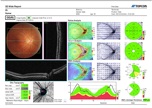
As a cataract and refractive surgeon performing a high volume of surgical procedures, including minimally invasive glaucoma surgery (MIGS), I need technology that can help me keep pace with my […]
IOTM: Melanotic Choroidal Nevus
IOTM: Melanotic Choroidal Nevus by Ranjit Dhaliwal, MD, FRCSC, FACS of The Retina Eye Center in Augusta, GA.
Efficiency in OCT Imaging: The Benefits of a Multi-Modality, Automated OCT
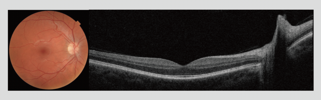
Efficiency is a primary practice management concern for today’s ophthalmology clinics because wasted time impacts patient satisfaction, frustrates doctors and staff, and leads to decreased revenues and profitability.
IOTM: Diabetic Traction, Macular Hole, Retinal Detachment
Image of the Month: Diabetic Traction, Macular Hole, Retinal Detachment by Leon Paolo Lara, MD.
Watch How This Top Practice Puts Customers First
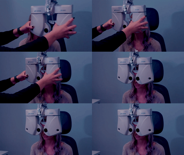
Dr. Shane Pressonn of Karns Vision in Knoxville, TN shares how Topcon’s technology and service has powered his clinic to drive efficiencies.
How to Capture a Radial Anterior Segment Scan with Maestro2
Watch how to capture a radial anterior segment scan on the Maestro2 Automated OCT/Fundus Camera.
The Importance of Image Quality in Accurate Diagnosis
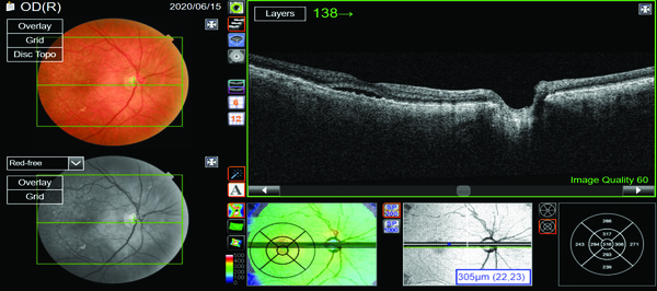
Imaging technologies play an important role in our profession, and the ability to visualize tissue and evidence of disease in detail is one of the cornerstones of our jobs.
















