Ciliary muscle dimensions by swept-source optical coherence tomography and correlation study in a large population

AUTHORS:

Fernández-Vigo JI, Shi H, Kudsieh B, Arriola-Villalobos P, De-Pablo Gómez-de-Liaño L, García-Feijóo J, Fernández-Vigo JÁ.

PUBLICATION:

Acta Ophthalmol. 2020 Jun;98(4):e487-e494. DOI: 10.1111/aos.14304

Study Purpose

To examine ciliary muscle (CM) dimensions in vivo by swept-source optical coherence tomography (SS-OCT) in a large healthy population.

Overview

Study Design

    Cross-sectional study

# of Eyes/
Patients

• 495 eyes from 495 subjects

Study Device

  • DRI OCT Triton™ (Topcon, Tokyo, Japan)

Outcome Measures

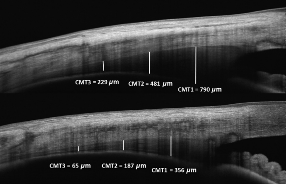
• CM length (CML), CM area (CMA) and CM thickness 1000 µm (CMT1), 2000 µm (CMT2) and 3000 µm (CMT3) from the scleral spur in the temporal and nasal quadrants. • Anterior chamber angle (ACA) and iris thickness 500 µm (IT500) from the scleral spur were also measured

Results

  • Mean CML was 4.57 ± 0.73 mm (range 2.16–6.97) and 4.45 ± 0.63 mm (2.42–6.37) in the temporal and nasal quadrants, respectively, with no differences detected between quadrants (p = 0.223).
  • Reproducibility of the CM measurements was excellent (ICC > 0.878)
  • Negative correlations were observed between refractive error and CML (R = -0.519; p < 0.001), and between refractive error and CM thickness (R ≥ -0.386; p < 0.001) or CMA (R = -0.538).
  • Negative correlations were found between CM dimensions (CML, CMT, and CMA) and age, with the strongest correlation observed for CML (R = -0.516; p < 0.001).
  • Ciliary muscle dimensions showed correlations with ACA (CML R = 0.499; CMT R = 0.434; CMA R = 0.545; all p < 0.001) and slightly with IOP (R = -0.175; p < 0.001) but no correlation was observed with IT500 (p ≥ 0.088)

IMOvifa reduced measurement time by

39%

FIGURE

FIGURE.

Swept-source optical coherence tomography ciliary muscle (CM) measurements made on a thick CM (upper image) and a thin CM (lower image). Adapted from Acta Ophthalmol. 2020 Jun;98(4):e487-e494. DOI: 10.1111/aos.14304

Conclusions

• This is the first study to use SS-OCT to assess CM dimensions in a large Caucasian population. • A SS-OCT instrument was used to measure CM dimensions with excellent reproducibility. • Data indicated a larger CB in myopia, a decrease produced with age, no correlation with gender, and good correlation with angle. • Limitations included, only assessing the horizontal quadrant, occasional difficulty determining the ciliary pigmented epithelium boundary, and the posterior zonules are not visible in the OCT images.


Product Comparison

Rotary Prism
Autorefraction

Keratometry

Auto Measure

Auto Right/Left

Full Auto
Align/Measurement

Rotary Prism
Autorefraction

Keratometry

Corneal Topography

Auto Measure

Auto Right/Left

Full Auto
Align/Measurement

Rotary Prism
Autorefraction

Keratometry

Auto Measure

Size and Weight

Height
XX Inches

Width
XX Inches

Depth
XX Inches

Weight
XX Pounds

Height
XX Inches

Width
XX Inches

Depth
XX Inches

Weight
XX Pounds

Height
XX Inches

Width
XX Inches

Depth
XX Inches

Weight
XX Pounds

Lorem Ipsum

X.X”

360° Rotating
Touchscreen

Control Lever

X.X”

LCD Screen Panel

Wavefront

Control Lever

8.5″

LCD Screen Panel

Lorem Ipsum

EMR

EMR Connectivity

Corneal Aberrometry

Ocular Aberrometry

Pupillometry

Tear Film Assessment

Viewer Software Available

EMR

EMR Connectivity

EMR

EMR Connectivity

The Topcon Healthcare Ophthalmology Solution

Lorem ipsum dolor sit, consectetuer adipiscing elit, sed diam nonummy.

The Topcon Healthcare Myopia Solution

Lorem ipsum dolor sit, consectetuer adipiscing elit, sed diam nonummy.

Fundus Cameras

Lorem ipsum dolor sit, consectetuer adipiscing elit, sed diam nonummy.

Fundus Cameras

Lorem ipsum dolor sit, consectetuer adipiscing elit, sed diam nonummy.