Dynamic Pupillary Response in Multiple Sclerosis Patients with and without Optic Neuritis

AUTHORS:

Gil-Casas A, Piñero DP, Molina-Martín A.

PUBLICATION:

Biomedicines. 2023 Dec 17;11(12):3332. doi: 10.3390/biomedicines11123332

Study Purpose

To assess different parameters of the pupillary response in Multiple Sclerosis (MS) subjects with and without previous optic neuritis (ON) using a pupillometry module integrated into a topographer device and to compare these results with those obtained in an aged-matched sample of healthy subjects

Overview

Study Design

    Prospective study

# of Eyes/
Patients

24 eyes of healthy age matched subjects (control group), 22 eyes of subjects with MS (MS group), and 13 subjects with MS with previous ON (MSON group)

Study Device

    MYAH topographer (Topcon EU, Tokyo, Japan)

Outcome Measures

Pupillary parameters: ratio pupil max/min; latency; velocity and duration; contraction and dilation; and amplitude of contraction

Results

  • The control group showed a relative maximum pupil size slightly higher (46.38 7.62%) than the MS (42.39 8.32%) and MSON group (41.82 8.24%), but they were still very similar (p > 0.05)
  • The amplitude of pupil contraction in the MSON group (40.71 6.73%) was reduced with respect to the control group (45.22 3.29%) (p = 0.01) but not compared to the MS group (43.75 5.02%) (p = 0.23)
  • When comparing the latency of pupil contraction between groups, the MSON group spent more time (0.35 ± 0.13 s) than the control group (0.26 ± 0.05 s) initiating the constriction reaction (p = 0.03)
  • In terms of the dilatation function, none of the analyzed parameters (velocity, latency, and duration) showed statistically significant differences between the three study groups. Even so, the speed of dilation was faster in the control group (0.89 ± 0.34 mm/s), with no statistically significant differences between the MS and MSON groups (0.74 ± 0.29 mm/s and 0.64 ± 0.26 mm/s, respectively)

IMOvifa reduced measurement time by

39%

FIGURE 1

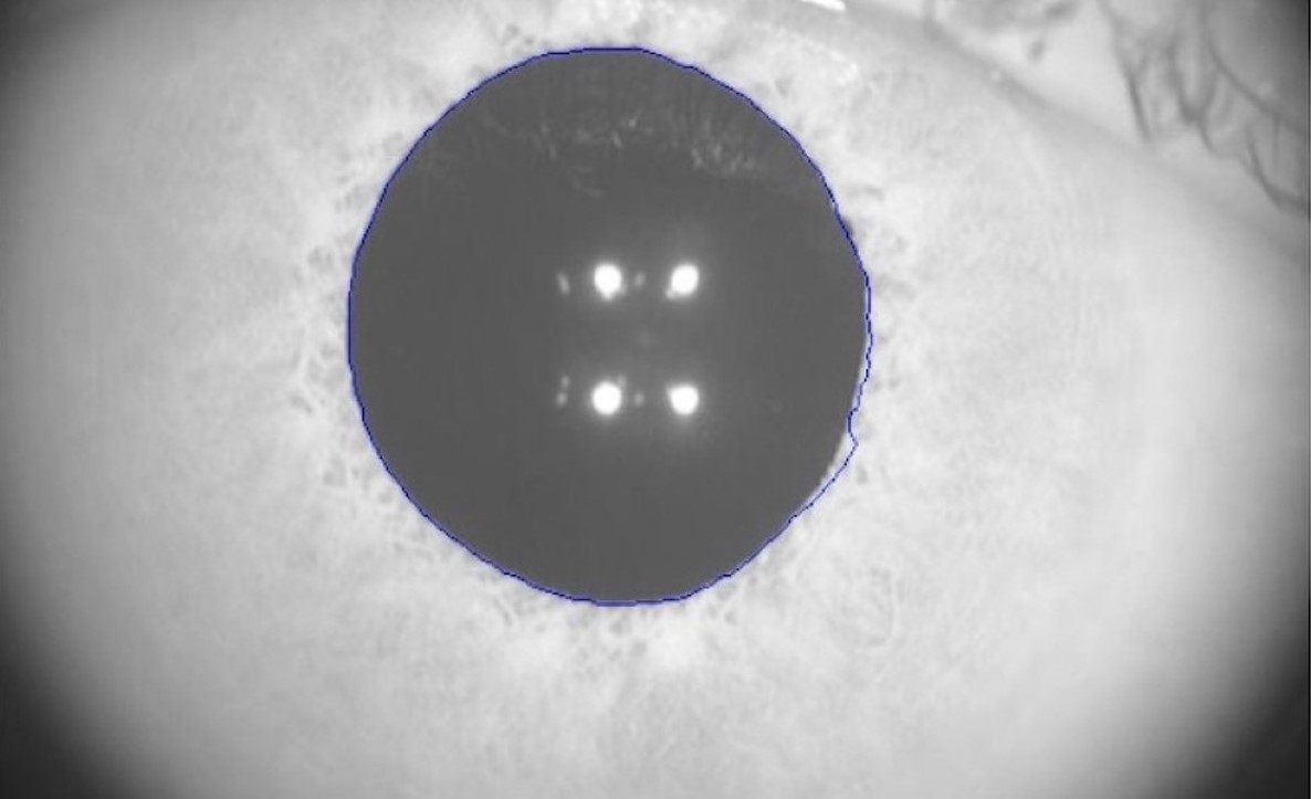
FIGURE 1.

Real image taken by MYAH topograher with the pupillometry module. The pupillary diameter is automatically detected and outlined in blue. Adapted from Biomedicines. 2023 Dec 17;11(12):3332. doi: 10.3390/biomedicines11123332

Conclusions

• The results obtained in the present paper showed that most of the pupillary parameters in MS subjects (without ON and normal CDVA) were comparable to those of healthy age matched controls • Based on the results of this study, the contraction process, especially latency and amplitude, were found to be affected in subjects with MS and previous ON


Product Comparison

Rotary Prism
Autorefraction

Keratometry

Auto Measure

Auto Right/Left

Full Auto
Align/Measurement

Rotary Prism
Autorefraction

Keratometry

Corneal Topography

Auto Measure

Auto Right/Left

Full Auto
Align/Measurement

Rotary Prism
Autorefraction

Keratometry

Auto Measure

Size and Weight

Height
XX Inches

Width
XX Inches

Depth
XX Inches

Weight
XX Pounds

Height
XX Inches

Width
XX Inches

Depth
XX Inches

Weight
XX Pounds

Height
XX Inches

Width
XX Inches

Depth
XX Inches

Weight
XX Pounds

Lorem Ipsum

X.X”

360° Rotating
Touchscreen

Control Lever

X.X”

LCD Screen Panel

Wavefront

Control Lever

8.5″

LCD Screen Panel

Lorem Ipsum

EMR

EMR Connectivity

Corneal Aberrometry

Ocular Aberrometry

Pupillometry

Tear Film Assessment

Viewer Software Available

EMR

EMR Connectivity

EMR

EMR Connectivity

The Topcon Healthcare Ophthalmology Solution

Lorem ipsum dolor sit, consectetuer adipiscing elit, sed diam nonummy.

The Topcon Healthcare Myopia Solution

Lorem ipsum dolor sit, consectetuer adipiscing elit, sed diam nonummy.

Fundus Cameras

Lorem ipsum dolor sit, consectetuer adipiscing elit, sed diam nonummy.

Fundus Cameras

Lorem ipsum dolor sit, consectetuer adipiscing elit, sed diam nonummy.