Evaluation of scleral thickness in patients with Fuchs endothelial dystrophy

AUTHORS:

Korkmaz I, Degirmenci C, Selver OB, Palamar M.

PUBLICATION:

Graefes Arch Clin Exp Ophthalmol 261, 2883–2889 (2023). PMID: 37178183 DOI: 10.1007/s00417-023-06107-z

Study Purpose

The aim of this study is to evaluate scleral thickness using AS-OCT (anterior-segment OCT) in patients with FED (Fuchs endothelial dystrophy) and compare the results with healthy individuals and to make an assumption of the affection of the sclera

Overview

Study Design

    Prospective cross sectional study

# of Eyes/
Patients

• 32 eyes of 32 patients with FED and 30 eyes of 30 age, gender, spherical equivalent and axial length matched healthy participants

Study Device

  • Anterior segment module of the SS-OCT Triton (AS-OCT, DRI Triton, Topcon, Japan)
  • Specular Microscopy (Nidek Co, Ltd, Gamagori, Japan)

Outcome Measures

• Scleral thickness in superior, inferior, nasal, and temporal quadrants 6mm, 4mm and 2mm posterior to the scleral spur – measured with AS-OCT • Endothelial cell density (ECD) and central corneal thickness (CCT) – measured with specular microscopy

Results

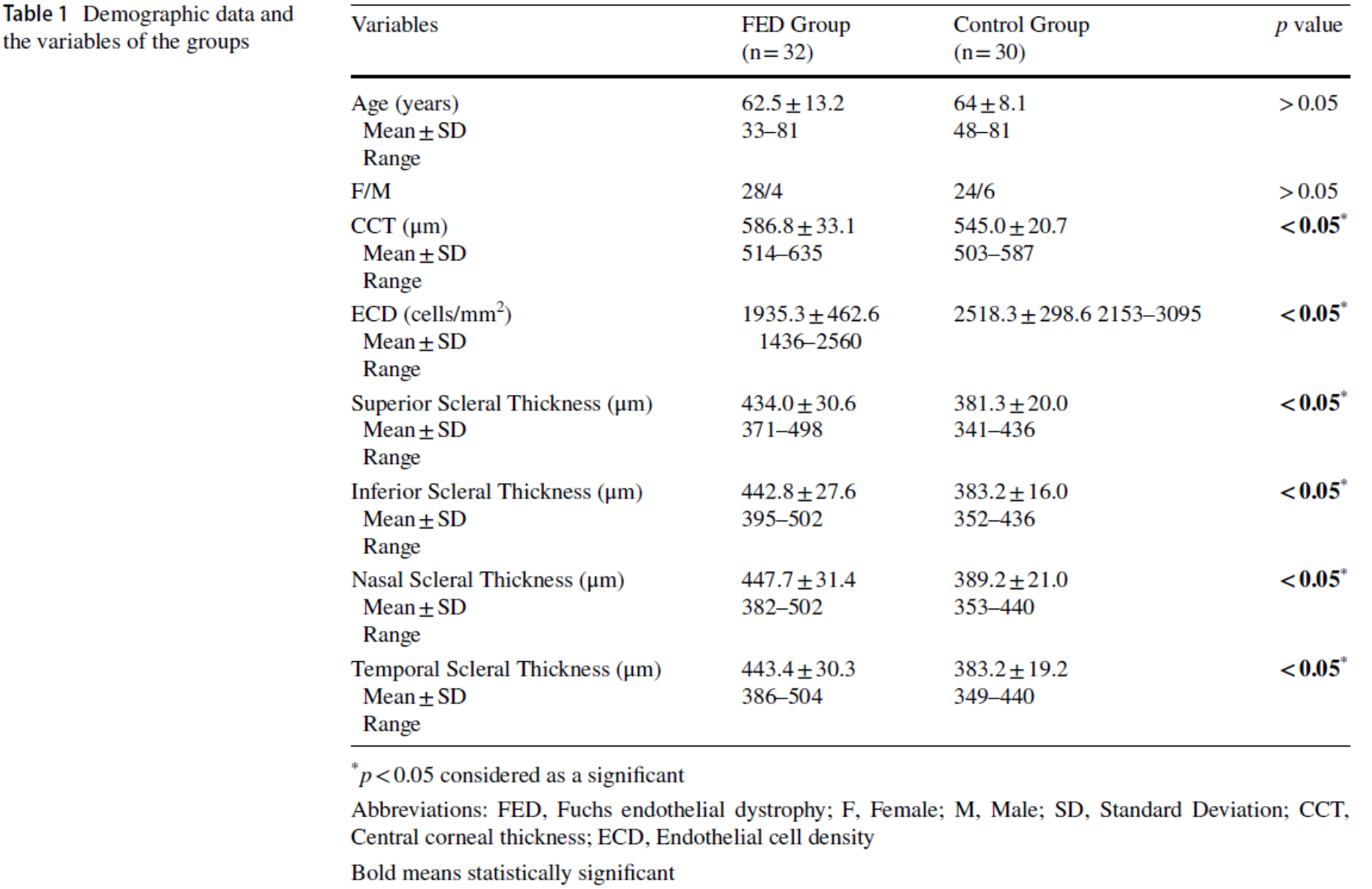
  • Mean CCT was significantly greater in the FED group (mean 586.8 ± 33.1; range 514–635 μm) than in the control (mean 545.0 ± 20.7; range 503–587 μm) (p = 0.000).
  • Mean scleral thickness 6 mm posterior to the scleral spur in the FED group was significantly higher in all quadrants than in the control group (p = 0.000) (Table 1).
  • Mean ECD was lower and mean CCT was higher in the FED group than in healthy individuals. (Table 1).
  • Mean scleral thicknesses at 2mm and 4mm posterior to the scleral spur in the FED group were significantly higher in all quadrants than in the control group (p=0.000).

IMOvifa reduced measurement time by

39%

TABLE 1

TABLE 1.

Adapted from Graefe’s Arch Clin Exp Ophthalmol (2023) 261:2883–2889 DOI: 10.1007/s00417-023-06107-z

Conclusions

• Scleral thickness, as measured by AS-OCT, was found to be significantly higher in patients with FED compared to the control group. These findings suggest that the accumulation of extracellular deposits in FED may not be limited to the cornea but may also affect the sclera.


Product Comparison

Rotary Prism
Autorefraction

Keratometry

Auto Measure

Auto Right/Left

Full Auto
Align/Measurement

Rotary Prism
Autorefraction

Keratometry

Corneal Topography

Auto Measure

Auto Right/Left

Full Auto
Align/Measurement

Rotary Prism
Autorefraction

Keratometry

Auto Measure

Size and Weight

Height
XX Inches

Width
XX Inches

Depth
XX Inches

Weight
XX Pounds

Height
XX Inches

Width
XX Inches

Depth
XX Inches

Weight
XX Pounds

Height
XX Inches

Width
XX Inches

Depth
XX Inches

Weight
XX Pounds

Lorem Ipsum

X.X”

360° Rotating
Touchscreen

Control Lever

X.X”

LCD Screen Panel

Wavefront

Control Lever

8.5″

LCD Screen Panel

Lorem Ipsum

EMR

EMR Connectivity

Corneal Aberrometry

Ocular Aberrometry

Pupillometry

Tear Film Assessment

Viewer Software Available

EMR

EMR Connectivity

EMR

EMR Connectivity

The Topcon Healthcare Ophthalmology Solution

Lorem ipsum dolor sit, consectetuer adipiscing elit, sed diam nonummy.

The Topcon Healthcare Myopia Solution

Lorem ipsum dolor sit, consectetuer adipiscing elit, sed diam nonummy.

Fundus Cameras

Lorem ipsum dolor sit, consectetuer adipiscing elit, sed diam nonummy.

Fundus Cameras

Lorem ipsum dolor sit, consectetuer adipiscing elit, sed diam nonummy.