To evaluate the use of non-mydriatic fundus photographs (NMFP) combined with OCT to facilitate ultra-rapid remote diagnosis and stroke alert for patients with acute vision loss presenting to the emergency department (ED)
Overview
Study Design
Prospective evaluation of all central and branch retinal artery occlusions (CRAO and BRAO) between 06/06/2023- 06/06/2024 who had NMFP-OCT imaging in Emory University’s general ED affiliated with a stroke center
# of Eyes/ Patients
26 patients diagnosed with retinal artery occlusions (22 CRAOs and 4 BRAOs) in ED
Study Device
Maestro2 NMFP-OCT (Topcon, Japan)
Outcome Measures
Time between vision loss onset and presentation to ED, time and results of ocular imaging, quality of ocular imaging, and number of patients undergoing thrombolysis treatment
Results
Five patients presented to ED within 4.5 hours of vision loss onset, six presented between 4.5 and 12 hours and 15 presented after 12 hours of symptom onset. On average, NMFP-OCT was performed within 141 minutes of presentation to ED (range 27-422 minutes)
Of the nine patients who underwent NMFP-OCT imaging within 12 hours of symptom onset, 5 had subtle retinal whitening on color photographs consistent with acute ischemia but all showed inner retinal hyperreflectivity/edema on OCT
Quality of color fundus photographs and OCT was good with average scores of 3.3/5 and 4.2/5, enabling diagnosis of acute CRAO or BRAO from 19 of 26 non-mydriatic color fundus photographs and 25 of 26 OCT images
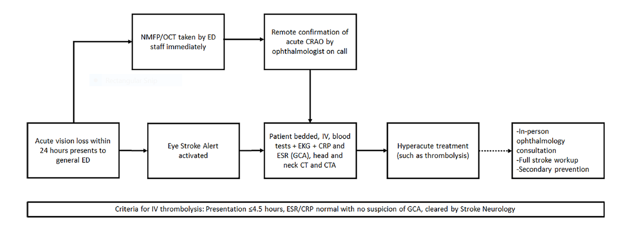
Diagnosis of acute RAO was made remotely with NMFP-OCT within 4.5 hours in 4 patients, 2 of whom received intravenous thrombolysis. All patients underwent a complete stroke workup while in the ED and were discharged home with appropriate secondary prevention of stroke (Figure 1)
IMOvifa reduced measurement time by
39%
TABLE 1
TABLE 1.
All patients presenting
to Emory’s general ED with acute
painless vision loss within 24 hours
are triaged to undergo NMFP-OCT
imaging immediately. Adapted from
J Stroke Cerebrovasc Dis. 2024 Jul
28;33(9):107895.
Conclusions
• Implementation of Maestro NMFP-OCT in a general ED enables rapid remote diagnosis of retinal artery occlusions and facilitates initiation of an eye stroke protocol in acute patients before in-person ophthalmology consultation is performed • OCT complements color fundus photography and provides greater diagnostic accuracy in hyperacute cases with near-normal appearing ocular fundi