Faricimab in neovascular AMD: first report of real-world outcomes in an independent retina clinic

AUTHORS:

Stanga PE, Valentin-Bravo FJ, Stanga SEF, Reinstein UI, Pastor-Idoate S, Downes SM

PUBLICATION:

Eye(2023) 37:3282-3289; PMID: 36959312 DOI: 10.1038/s41433-023-02505-z

Study Purpose

Present week four clinical outcomes from a real-world study in an independent specialized retina clinic to assess the safety, efficacy, and durability of faricimab administered as customized treatment in patients with nAMD.

Overview

Study Design

    Observational, retrospective

# of Eyes/
Patients

• 11 eyes from 9 patients

Study Device

  • DRI Triton SS-OCT (Topcon, Tokyo, Japan)
  • Optos California, Silverstone (Optos PLC; Dunfermline, Scotland, United Kingdom)

Outcome Measures

• Anatomical and functional features at baseline and 1 month after faricimab intravitreal injection

Results

  • At one month following the first faricimab intravitreal injection, four eyes (36.36%) gained at least one line of vision, six eyes (54.54%) remained stable, and one eye (9.09%) lost one line of vision.
  • Overall average CRT at baseline was 565.41 +/- 391.38 µm which improved significantly to 326.97 +/- 226.48 µm at one month.
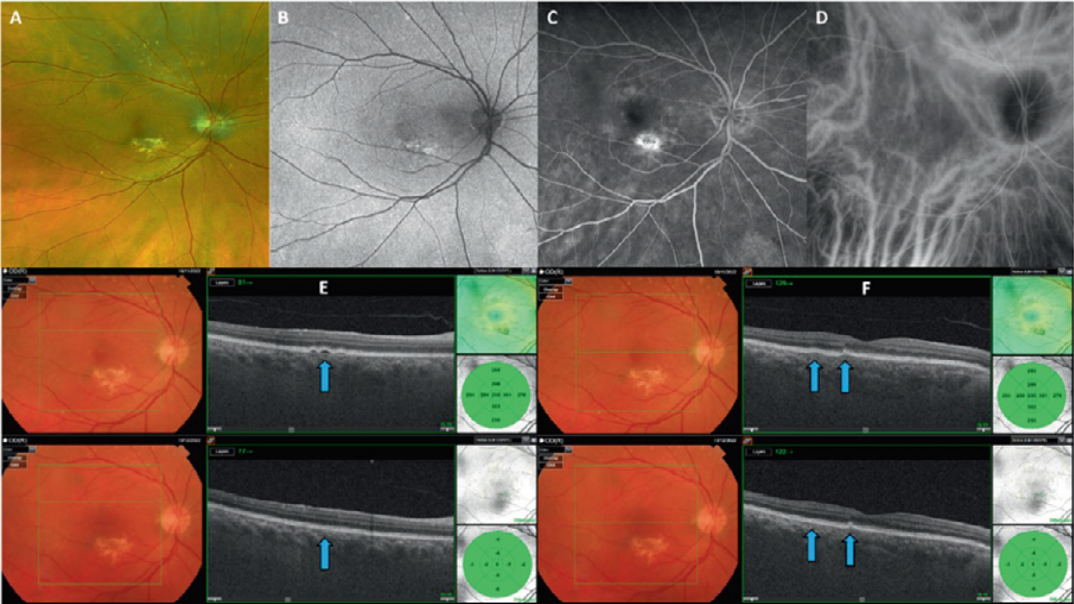
  • PEDs were present in eight eyes (72.72%) at the initial examination and morphology changes such as flattening were observed in all of them 1 month after the first faricimab injection (Figure).

IMOvifa reduced measurement time by

39%

FIGURE

FIGURE.

Resolution of multiple PEDs after the first faricimab intravitreal injection. Case 9: Patient diagnosed with nAMD using multimodal and simultaneous imaging including UWF RG (A), FAF (B), FFA (C ) and ICGA (D), with navigated SSOCT. In E and F, a resolution of multiple PEDs was observed after the first injection of faricimab (blue arrows) Adapted from Eye(2023) 37:3282-3289; PMID: 36959312 DOI: 10.1038/s41433-023-02505-z

Conclusions

• Real-world results with faricimab in terms of improvement of BCVA and resolution of subretinal fluid or subretinal pigment epithelium haemorrhage are similar or even better when compared to other licensed anti-VEGF treatments at one month after the first injection. • A way forward would be to identify the response therapy identifying specific OCT ‘biomarkers’ that will enable the identification of which treatment regimen is the most appropriate for each patient. • A personalized medicine approach would be more cost –effective and reduce the number of appointments and IVI needed per year. • Study limitations: Small sample size and short follow-up. Only 3 treatment naïve eyes

Product Comparison

Rotary Prism
Autorefraction

Keratometry

Auto Measure

Auto Right/Left

Full Auto
Align/Measurement

Rotary Prism
Autorefraction

Keratometry

Corneal Topography

Auto Measure

Auto Right/Left

Full Auto
Align/Measurement

Rotary Prism
Autorefraction

Keratometry

Auto Measure

Size and Weight

Height
XX Inches

Width
XX Inches

Depth
XX Inches

Weight
XX Pounds

Height
XX Inches

Width
XX Inches

Depth
XX Inches

Weight
XX Pounds

Height
XX Inches

Width
XX Inches

Depth
XX Inches

Weight
XX Pounds

Lorem Ipsum

X.X”

360° Rotating
Touchscreen

Control Lever

X.X”

LCD Screen Panel

Wavefront

Control Lever

8.5″

LCD Screen Panel

Lorem Ipsum

EMR

EMR Connectivity

Corneal Aberrometry

Ocular Aberrometry

Pupillometry

Tear Film Assessment

Viewer Software Available

EMR

EMR Connectivity

EMR

EMR Connectivity

The Topcon Healthcare Ophthalmology Solution

Lorem ipsum dolor sit, consectetuer adipiscing elit, sed diam nonummy.

The Topcon Healthcare Myopia Solution

Lorem ipsum dolor sit, consectetuer adipiscing elit, sed diam nonummy.

Fundus Cameras

Lorem ipsum dolor sit, consectetuer adipiscing elit, sed diam nonummy.

Fundus Cameras

Lorem ipsum dolor sit, consectetuer adipiscing elit, sed diam nonummy.