- MAESTRO
The development of a reference database with the Topcon 3D OCT-1 Maestro
AUTHORS:
Chaglasian M, Fingeret M, Davey PG, Huang WC, Leung D, Ng E, Reisman CA*
PUBLICATION:
Clin Ophthalmol. 2018 May 7;12:849-857. doi: 10.2147/OPTH.S155229.
Study Purpose
To report the range of thickness values for the new Topcon Maestro 3D OCT device with 2 scan size settings: the 12×9 mm wide field and 6×6 mm scans
Overview
Study Design
- Prospective, multicenter cohort study
# of Eyes/
Patients
399 healthy eyes of 399 subjects
Study Device
- Topcon 3D OCT-1 Maestro
Outcome Measures
Average and 1st, 5th, 95th, and 99th percentile ranges for OCT parameters: Early Treatment Diabetic Retinopathy Study macula full retinal thickness (FRT), ganglion cell + inner plexiform layer thickness (GCL + IPL), ganglion cell complex (GCC) thickness, circumpapillary retinal nerve fiber layer (cpRNFL) thickness
Results
- The mean age was 46.3 ± 16.3 years (ranged 18–88 years) and 57% of the subjects were female
- Eighteen percent of the subjects were Hispanic or Latino. Fifty-nine percent of the subjects were Caucasians, followed by Black/African American (20%), Asian (13%), Native American/ Pacific Islander (2%), and American Indian/Alaskan Native (1%)
- The mean foveal FRT was 237.079 ± 20.899 µm for the 12×9 mm scan and 234.000 ± 20.657 µm for the 6×6 mm scan; FRT reduced with age in the outer ETDRS quadrants
- The mean GCL + IPL thickness was 71.363 ± 5.924 µm for the 12×9 mm scan and 71.726 ± 5.880 µm for the 6×6 mm scan; a reduction in GCL + IPL thickness was seen with age
- The mean GCC thickness was 105.949 ± 8.533 µm for the 12×9 mm scan and 106.698 ± 9.094 µm for the 6×6 mm scan; overall, GCC thickness decreased with age
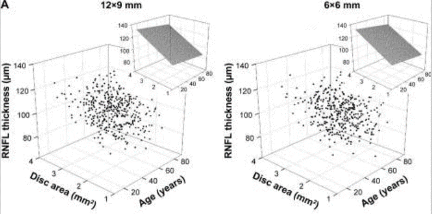
- The mean cpRNFL thickness was 104.720 ± 11.829 µm for the 12×9 mm scan and 104.036 ± 11.341 µm for the 6×6 mm scan; age had little or no effect, whereas disc area showed large degree of influence on the cpRNFL thickness (Figure 1)
IMOvifa reduced measurement time by
39%
FIGURE 1

FIGURE 1.
The relationships of cpRNFL thickness with age and disc area displayed in the 3D scatter plots. Adapted from Clin Ophthalmol. 2018 May 7;12:849-857.doi: 10.2147/OPTH.S155229
Conclusions
Across all parameters, the measurements obtained by the 12×9 wide, the 6×6 macula, and 6×6 disc scan modes on the Topcon 3D OCT-1 Maestro were generally similar and their trends were consistent with those in the literature. The reference limits at the 1st, 5th, 95th, and 99th percentile points establish thresholds to provide for the quantitative comparison of the RNFL, optic nerve head, and the macula in the human retina to a database of known healthy eyes.